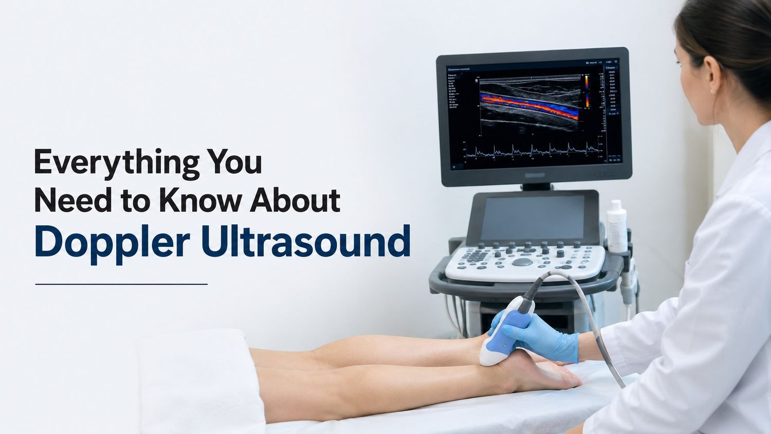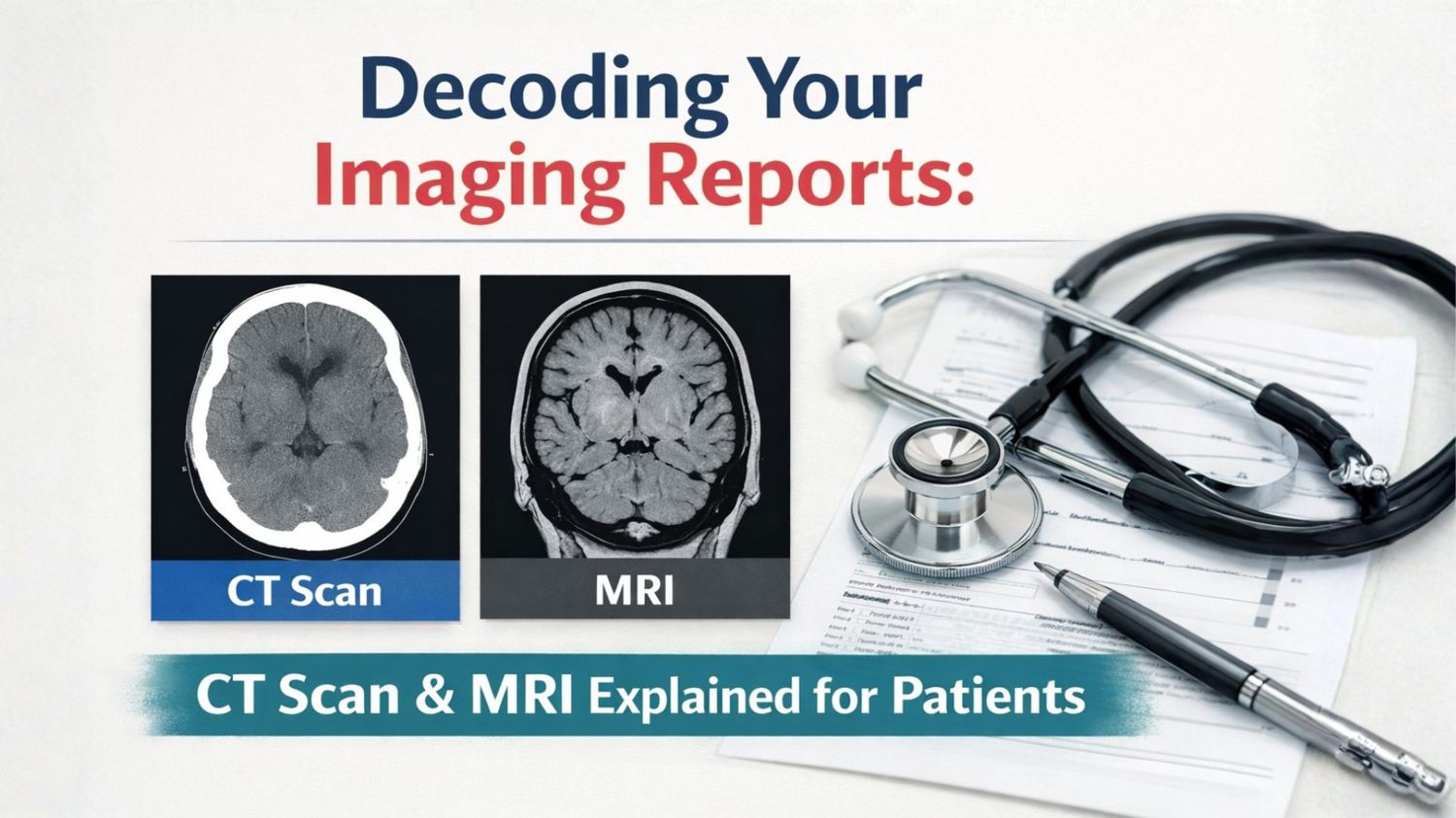
The powerful tools which help the doctors to see what is inside your body without surgery are known as Medical imaging tests like CT scans and MRIs. The complicated medical terminology is involved in these results of the scans and often they seem to be confusing to the patients. Thus understanding of the basics of the reports will help the patient more confident and informed about the health of the patient. An MRI scan is a medical imaging technique that uses strong magnetic fields and radio waves to produce detailed images of the body’s internal structures. Soft tissues such as the brain, spinal cord, muscles, ligaments, and internal organs are usually examined by this technique. In the case of emergencies, MRI scan is widely used due to its detailed evaluation of the soft tissue injuries and CT scan is used because of the speed of the scan.
MRI Scans
MRI scans are usually recommended to the patients because these MRI scan images help the doctor to examine the detailed images of the tissues and structures inside your body. Thus these scans results are helpful for the accurate diagnosis. MRI scans aid drastically where the symptoms shows the condition is related to nerves, joints, or internal organs since the images produced by the MRI are of high resolution and clarity.
MRIs are useful for the following:
- Muscles
- Ligaments
- Joints
- Brain
- Spinal cord
- Tumours
- Neurological conditions
- Organs like the liver, kidneys, and heart
The MRI scans are safe and non-invasive and they require longer time than a CT scan.
CT Scans
CT stands for Computed Tomography and these scans are used to create the detailed cross-sectional images of the body using the X-rays. The technique makes the use of the multiple X-ray images taken from different angles and through the help of the computer it produces the clear and magnified images of the organs, tissues and bones. These scans are quick at detecting tumours, identifying internal injuries, guiding treatment decisions in emergency situations and in diagnosing the vascular conditions.
CT scans are used to detect:
- Bone injuries
- Fractures
- Tumours
- Cancers
- Infections
- Internal bleeding
- Lung
- Chest conditions
In a CT scan a contrasted dye may be taken orally or injected in order to highlight the areas and making the diseased or abnormalities easier to detect.
In order to highlight certain areas during the scan a contrasting agent is used, iodine-based contrast is commonly used in the CT scans whereas in the MRI gadolinium-based contrast are used. These substances help to distinguish between normal and abnormal tissues and provide the images with more clarity and high resolution.
Main Difference between an MRI and CT scan
MRI:-
- Better for viewing of soft tissues, fat, water, and muscles.
- Takes more time for the images.
- More expensive.
- Uses strong Magnetic field.
- Can cause claustrophobia.
- Longer (typically 20 to 90 minutes.
CT Scan:-
- Optimal for viewing bones, such as the spine, and veins clot.
- Produce images faster.
- Less expensive.
- Uses ionizing radiations.
- Do not cause claustrophobia.
- Faster (typically 5 to 10 minutes).
Risks of the MRI Scan
- Insulin pumps
- Pacemakers
- Cochlear implants
- Implanted cardiac defibrillator
- Deep brain stimulators
- Valgus nerve stimulators
Uses of CT Scans
CT scans are versatile in nature, and their uses include the following diseases diagnosis:
- Abscesses within different body areas
- Cancer screening and diagnosis
- Bladder and kidney stones
- Coronary artery diseases
- Blood clots
- Sinusitis
- Internal injuries from trauma
- Bone injuries
- Spine injuries
Safety Considerations and Risk Management during the CT Scan and MRI scan
Following are the safety considerations during the CT & MRI scans:
- The principle of ALARA (As Low As Reasonably Achievable) is used during the scan.
- The diagnosis quality is maintained while reducing and minimising the risk of the radiations.
- The radiation exposure remains within the safe limits.
- Magnetic field effect is used instead of the radiations.
Understanding the Imaging Reports
The imaging reports are written by the trained Radiologist who is specialised in interpreting the medical images obtained through the MRI and CT scans. The reports include the following:
- Name of the patient
- Age of the patient
- Date of the scan
- Type of scan
- Reason for the test
- Common observations obtained through the images
- Summary of the report
- Diagnosis based on the findings of the report
Clinical Indication section of the Imaging Report
This section shows the clinical question your physician had when requesting the scan. Thus this part is most focused where answers to more of the questions is answered and helped by the results interpretation of the images and it guides the radiologist’s interpretation of the images of your scan.
Findings section of the Imaging Report
This is the most detailed section of the imaging report, and it lists the common observations from the scanned images. The descriptions of the common abnormalities are listed in this section.
Common Findings in the Reports
- If any mass in the form of cancerous cells is there or not
- If inflammation is there due to any sort of swelling or injury
- Lesions
- Fractures if any reported during the scans
Comparison section of the Imaging Report
This section of the report makes the comparison of the patient’s images results with its past findings and results if any and this section can also be deleted if no prior scan images are available with the lab and thus it helps to see the changes over a period of time between two successive scans and it help doctors track progress or detect new issues. Thus it is helpful for tracking a condition or follow-up after treatment.
Impression section of the Imaging Report
The most critical part of the imaging report is the impression section of the report in which the summary of the ley observations from the findings section are summarised by the radiologist and explains the interpretation of the common findings and answers the common questions posed in the clinical indication section. A diagnostic assessment is usually given in this section.
Common Terminology of the Report
What the radiologist observed in your report is described by the key imaging terminology or words and this helps the patients to read the imaging report. In the Findings and Impression sections these terms are frequently used and help present clinical imaging findings clearly and accurately.
General Report Words
Normal or unremarkable:- Nothing unusual was observed in the report
Incidental Finding:- Result that was not related to the reason why the particular scan was carried out
Follow up needed :- Another imaging test is recommended by the radiologist in order to analyse the situation more clearly
Impression or summary:- Interpretation of the results by the radiologist
Comparison :- Comparison of the patient’s images results with its past findings
Comparison
Comparison of the patient’s images results with its past findings
Thus these are the some of the methods through which the decoding of your imaging reports can be done easily and it will help the patients to make more informed decisions after analysing the results of the imaging reports by their health care providers and control the treatment process and diagnosis of the particular abnormality on time and help the patients to recover faster without wasting much of the important time.