Add Member
Provide email & phone to receive medical reports and notifications.
Blogs

Health Insights & Medical Updates

Stay informed with expert health tips, diagnostics knowledge, and latest lab updates.

Latest News
Healthcare
What is Mammography? A Complete Guide for Women in Bang...

The specialized medical technique that uses low-dose X-rays to examine the breast tissue is known as Mammography. The changes that are not visible or felt during the physical examination are detected in a mammogram and this is the primary purpose of a mammogram. A mammogram can detect and identify the changes such as small lumps, calcifications, and other abnormalities before even the symptoms develop and seen. Early detection significantly increases the chances of successful treatment and recovery, making mammography an important screening tool for the detection breast cancer at an early stage.Regular screening plays a very important role in the early detection and screening of the breast diseases and thus for the overall well-being of the breast health of the women it is one of the most effective and important diagnostic tool available today. Understanding mammography can help make informed decisions about preventive healthcare for the women in cities like Bangalore where the access to advanced healthcare facilities is readily available.Presently worldwide, breast cancer is one of the most common cancers affecting women today. In India especially in the urban areas the cases of the breast cancer have been increased and in most of the cases women during the early stages of breast cancer, do not experience symptoms of the breast cancer and that is why routine screening becomes essential for the women and is very helpful.Types of MammographyFollowing are the main types of Mammography:Screening Mammography: It is performed on the women who do not have any noticeable breast symptoms.Diagnostic Mammography: When a woman experiences the symptoms such as breast lump, breast pain, nipple discharge etc. the diagnostic mammography is performed.Digital Mammography: The breast images are captured electronically instead of using the traditional films.3D Mammography (Tomosynthesis): From the different angles the multiple breast images are captured and it combines them into a 3-D pictures.Procedure of MammographyMedical gown is usually recommended during the entire process.On the flat imaging platform, the breast is placed.The breast is allowed to evenly spread on the compression plate so that the tissues can spread evenly for the mammogram.From the different angles the X-ray images are taken.Similar process is repeated with other breast as well.The entire procedure takes about 15-30 minutes to complete.Is Mammography Safe?Mammography is considered the safe procedure and the amount of the radiations used is also very low and thus it is considered safe as per the medical safety standards.The benefits of early cancer detection greatly outweigh the minimal risks associated with radiation exposure.While producing highly detailed images the modern digital mammography systems use even lower radiation doses.Mammogram is for:Women who should undergo mammography are:Whose age is 40 years and above.Having the family history of breast cancer.Those who have the genetic mutations associated with breast cancer risk.Those who have the breast abnormalities earlier.Women experiencing breast-related symptoms Uses of MammographyMammography helps in:Detection of the breast cancer at an early stage.Identification of the non- cancerous breast conditions.Monitoring the changes in the breast tissues over time.Through the early intervention the reduction in the breast cancer related mortality.Assisting doctors in planning appropriate treatment Benefits of MammographySome major benefits of Mammography include:Breast cancer’s early detection.Increased success rates for the treatment.Less treatment requirements.Survival rates improved.Detection of abnormalities before symptoms appear. Better long-term breast health monitoring. Limitations of MammographyMammography is not perfect but it is highly effective and some of its limitations are:Abnormalities are harder to detect because of the dense breast tissues.Due to false-positive results it may lead to additional testing issues.Occasionally the small cancers are missed.Sometimes additional imaging may be required necessary. ConclusionWe can conclude from above that women over 40 or those with higher risk factors, can make a substantial difference in breast health and in the early detection and diagnosis of breast cancer Mammography plays a vital role. Regular screening helps in the early detection and helps to save many lives. In Bangalore women have the access to the advanced mammography services which makes the preventive healthcare more convenient than ever. The women can take the proactive steps towards maintaining their breast health and overall well-being. The breast screening packages are offered by the leading hospitals and diagnostic centres which includes clinical breast examinations, mammography, and specialist consultations.

Healthcare
Everything You Need to Know About Doppler Ultrasound

Doppler Ultrasound is the non-invasive technique which uses high-frequency sound waves to evaluate blood flow through the body's blood vessels. The Doppler ultrasound measures the movement of the blood within arteries and veins unlike the standard ultrasound which creates the images of the tissues and the organs. It is widely used in the diagnosis conditions related to pregnancy, heart issues and blood vessels. Diagnosis of various vascular and cardiac conditions can be determined easily through this ultrasound as it helps to determine the abnormalities in blood circulation.It is widely used in the hospital and diagnostic centers to assess the blood flow in the arteries, determine the blockages and monitor the pregnancy and helps to evaluate the heart health.Preparation for a Doppler UltrasoundThe preparation for the tests includes the following requirements such as:Loose clothing is usually recommended for the patients for smooth processing.Fasting is usually recommended by the doctor for patients if it involves the abdominal blood vessels.For any medications if any please inform your doctor in advance.Staying hydrated is recommended during the ultrasound.Avoid applying the lotions on the area being scanned.Provide the medical information to your doctor before starting the procedure.Risks involved: Although it is the safest technique and is painless as well. Till date there are no known harmful effects associated with the procedure when performed by trained professionals. No discomfort is seen in the patients except the slight pressure from the transducer. Thus there are no major side effects of the ultrasound and in most of the cases the normal activities can be resumed immediately after the test.Advantages of Doppler USG ScansDoppler ultrasound has the following advantages such as:Non-Invasive technique.Radiation-Free procedure and is safe.Real-Time Results are obtained easily.Versatile techniqueCost-Effective in nature.Early Detection of the abnormalities is easy.Guidance for Treatment is provided through the assessments.Types of Doppler UltrasoundSpectral Doppler Ultrasound: It displays the flow of blood in the body in the form of the graphs and thus allows the doctors to measure the velocity of the blood flow.Colour Doppler Ultrasound: The direction and the speed of the blood flow is determined using the colour coding.Power Doppler: It is more sensitive than the colour Doppler and it can detect the slower blood flow as well.Duplex Doppler Ultrasound: It makes the use of the traditional ultrasound technology with the Doppler technology.Continuous Wave Doppler: It is quite useful for measuring high-speed blood flow and it continuously sends and receives the sound waves.General Procedure for Doppler UltrasoundThe general procedure for Doppler Ultrasound Scans are as follows:The patient is asked to lie down on the examination table before the ultrasound.The probe is placed by the radiologist on the concerned area.The blood flow is measured by the pitch of the sound.Graphs and pictures are created on the screen by the machine.High frequency sound waves are used by the radiologists.The radiologist may ask you to change positions during the procedure to get clearer images from different angles for the specific areas.The entire procedure takes around 20-45 minutes.The gel is wiped out of the skin once the scan is completed.Under what conditions can Doppler ultrasound help diagnosisDoctors recommend the Doppler ultrasound to diagnose:Narrowed arteries or veins.Blood clots, including deep vein thrombosis (DVT).Blood vessel injuries.Chronic venous insufficiency (CVI).Evaluate blood supply to a transplant organ like kidneys and pancreas.Tumours in blood vessels.Conclusion Thus it can be concluded that Doppler Ultrasound has wide applications and benefits in the field of medical science and is one of the most useful and non-invasive technique since the exposure to radiations is not there it is completely safe for the pregnant women, children and old age people as well. The extensive applications of this technique makes it so useful along with its cost effective advantage and the interpretation of the results is so easy which further helps in the diagnosis of the treatment after the detection of the abnormalities.

Healthcare
Doppler Ultrasound Test: Purpose, Procedure, Benefits &...

Doppler Ultrasound is widely used to evaluate blood flow in various parts of the body and it is one of the most essential diagnostic tool in the field of Medical science. This non-invasive procedure provides invaluable insights into your overall health by diagnosing the vascular conditions and other routine monitoring of the body. Doppler scans are considered more dynamic than the routine ultrasonography in which the pictures are collected over the computer screen whereas in the Doppler scans the measurement of the blood movement in vessels takes place in order to provide information such as the speed and direction of blood flow.Doppler scans help identify conditions like deep vein thrombosis (DVT) or peripheral artery disease (PAD) in a vascular type of medicine.They assess blood flow through the heart and its valves in the cardiology.Doppler ultrasound is used to monitor blood flow to and from the placenta, monitoring the health of a fetus.The procedure of the Doppler ultrasound takes about 15-45 minutes based on the area under examination and is comparatively safe even for the pregnant women since it does not use radiations. Doppler provides dynamic data, making it crucial for diagnosing and monitoring conditions affecting the cardiovascular system.Who might need a Doppler ultrasound?Patients with following conditions might need the Doppler ultrasound which are as follows:If the patient is Diagnosed with blood vessel-related disorder.If he has undergone with surgery, and want to check the blood flowIn pregnancy a colour Doppler Ultrasound is required.Purpose of the Doppler UltrasoundFollowing are the main areas where Doppler ultrasound is widely used and has extensive applications as well, such as:It helps to evaluate the circulation of the blood inside the body.To detect any sort of the blockages such as narrowed or blocked blood vessels in the body.Helps to access the vascular heath like the Deep Vein Thrombosis (DVT).It can assess and check the foetus health as well during the pregnancy.Helps to plan and monitor treatments such as vascular surgeriesUnder the following conditions as well the Doppler Ultrasound can be recommended as such:Leg pain or swelling Suspected blood clots Varicose veins Dizziness Stroke symptoms Poor circulation in the arms or legs Unexplained vascular abnormalitiesProcedure of the Doppler UltrasoundDoppler ultrasound uses the Doppler effect.In this procedure the pitch is changed by the sound waves when they are send into your body.The sound waves bounce off from the body’s moving red blood cells usually in the blood vessels it goes back to the probe to be detected.The direction of blood flow and how fast the blood is moving is also determined by the ultrasound.The sound waves travel during the ultrasound through the probe.The machine sends the high-frequency sound waves in the body.The change in the waves is examined by the probe.The gel is then wiped out of the body.Benefits of Doppler Ultrasound The benefits of the ultrasound are as follows:Radiation free technique and is painless.It is the safe technique.The precise and clear images are obtained in this process.Patient safety is considered as the priority.Transparent rates for the ultrasound.The detailed explanations during the results analysis are provided.Quick and Convenient to use.The results obtained are real time.Cost effective technique.Helps to access various health conditions.Safer during pregnancy as well.Understanding the ResultsThe results of the Ultrasound are easy to evaluate through the findings of the doctors and the Abnormal findings during the ultrasound may reveal:Blood clots Narrowed arteries Blocked blood vessels Poor circulation Heart valve abnormalities Reduced blood flow to organs Pregnancy-related circulation issues Based on the findings of the results the treatment and further assessment of the diagnosis process can be carried out under the guidelines of the concerned and trained doctors or sonographer.Conclusion Doppler Ultrasound is the most invaluable tool in the field of the medical science and it is the most safer, painless and non-invasive technique in nature. It helps to access the flow of blood inside your body and helps to determine lot of abnormalities such as circulatory and vascular conditions inside the body and most importantly it is considered safe during pregnancy as well where the foetal health can also be determined easily. The procedure is straightforward and offers a wealth of benefits for your health.

Healthcare
Doppler Ultrasound Explained: Uses, Benefits and How It...

The non –invasive imaging technique that uses sound waves to evaluate blood flow through the body's blood vessels. In this technique the red blood cells present in the body bounces back the sound waves in this type of the ultrasound and is known as Doppler Ultrasound. The change in the sound waves pitch can be easily seen on the computer screen by the healthcare provider. Thus this type of ultrasound helps to determine the blood flow direction and movement in the body and it also measures the blood movement within the arteries and veins. Diagnosis of various vascular and cardiac conditions can be determined easily through this ultrasound as it helps to determine the abnormalities in blood circulation.It is widely used in the hospital and diagnostic centers to assess the blood flow in the arteries, determine the blockages and monitor the pregnancy and helps to evaluate the heart health. It is the painless, safe and non-invasive technique since it does not use the radiations and thus it is safe for age groups including children and pregnant women as well.Preparation for the Doppler UltrasoundFasting is usually recommended before the ultrasound irrespective of the type of the ultrasound and reason for which it is carried out.Smoking should be avoided or stopped permanently.Consumption of the nicotine-containing products for a minimum of two hours before the test should be stopped.Procedure for the Doppler UltrasoundImaging technology is used by the sonographer or the specialist to carry out the ultrasound.Depending on the type of the ultrasound the position of the body is decided for the test.A gel is applied on the skin where the ultrasound is to be carried out.The sonographer glides a tiny probe on your skin in order to carry out the ultrasound.The probe helps the sound waves to travel during the ultrasound.The high-frequency sound waves are sent by the machine in the body.The waves bounce off the blood cells, and the pitch changes.The change in the waves is examined by the probe and then the graphs or pictures are converted by the machine.The gel is then wiped out of the body.A computer processes these signals and displays images and blood flow information on a monitor. Types of Doppler UltrasoundSpectral Doppler Ultrasound: It displays the flow of blood in the body in the form of the graphs and thus allows the doctors to measure the velocity of the blood flow.Colour Doppler Ultrasound: The direction and the speed of the blood flow is determined using the colour coding.Power Doppler: It is more sensitive than the colour Doppler and it can detect the slower blood flow as well.Duplex Doppler Ultrasound: It makes the use of the traditional ultrasound technology with the Doppler technology.Continuous Wave Doppler: It is quite useful for measuring high-speed blood flow and it continuously sends and receives the sound waves.Uses of Doppler UltrasoundDoppler ultrasound uses are as follows:It helps to determine the blood clots.Narrowed or blocked arteries can be detected easily.The condition of the heart can be determined.Doppler ultrasound evaluates blood flow between the mother, placenta, and fetus during the pregnancy.Helps to identify the damaged veins.Helps to evaluate blood flow to organsBenefits of Doppler UltrasoundDoppler ultrasound offers numerous advantages which are as follows:It is the non-invasive procedure.Radiation free imaging technique.Quick and Convenient to use.Real time results are obtained.Cost effective in comparison of other techniques.Provides valuable diagnostic information.The technique is safe during pregnancy.It is widely used for fetal monitoring and maternal health assessments.Conclusion Thus we can conclude that Doppler ultrasound is a valuable diagnostic imaging technique that helps healthcare providers assess blood flow throughout the body. It can detect clots, arterial blockages, heart conditions, circulatory disorders, and pregnancy-related complications by using the sound waves. It is one of the most widely used diagnostic tools in modern medicine due to its non-invasive nature, affordability and the real time imaging results provided by the ultrasound. Doppler ultrasound provides critical information that helps doctors make accurate diagnoses and develop effective treatment plans.

Healthcare
What is a Doppler Ultrasound? Uses, Benefits, Procedure...

A non-invasive diagnostic imaging test that uses high-frequency sound waves to evaluate blood flow through the body's blood vessels is known as Doppler Ultrasound. The Doppler ultrasound measures the movement of the blood within arteries and veins unlike the standard ultrasound which creates the images of the tissues and the organs. This helps doctors identify abnormalities in blood circulation and diagnose various vascular and cardiovascular conditions. The Doppler ultrasound is used widely because it is the painless, safe, radiation free and highly effective technique which helps in accessing the blood flow in different parts of the body. It is widely used in the diagnosis conditions related to pregnancy, heart issues and blood vessels.Types of Doppler ultrasoundsThe different types of Doppler ultrasounds include:Colour Doppler: The direction of the blood flow is detected by the changes in the sound waves into different colours as shown by the computer.Spectral Doppler: Graphical representation of blood flow over time.Duplex ultrasound: The width of the blood vessels can be checked using this type of the ultrasound and it combines the traditional ultrasound pictures with Doppler ultrasound and it also helps to show the blockages.Power Doppler: This test is used to show even very slow blood flow and is used to shows the presence of blood flow whereas the direction of the blood flow is not shown by this type of the ultrasound and is widely used to study blood flow inside organs.Transcranial Doppler ultrasound: This type of the Doppler ultrasound is used to detect strokes or subarachnoid haemorrhages and determines the blood flow in your brain.Procedure of the Doppler UltrasoundDoppler ultrasound uses the Doppler effect where the pitch is changes by the sound waves when they are send into your body.As and when the sound waves bounce off from the body’s moving cells in the blood vessels it goes back to the probe to be detected.In turn the auditory and visual representations of the blood flow is determined by the information through the Doppler effect.The direction of blood flow and how fast the blood is moving is also determined by the ultrasound.         This information provides information about:How fast or slow blood is moving and the actual condition of your blood circulation.Obstructions in the blood flow.Blood going in the wrong direction or pooling in a blood vessel.Why Doppler Ultrasound is used?No needles or surgical procedures are involved and thus it is the non-invasive technique.For all the age groups this process is safe and completely painless.It provides detailed insights into vascular health.The results are very quick and the entire process is very cost effective.Early detection of the diseases is important and it provides the real time images of the body parts under examination.Common Uses of Doppler UltrasoundDoppler ultrasound is used in a variety of medical conditions and it has following common uses:Blood Clots detection.Narrowed arteries and blocked diagnosis can be determined easily.Evaluating Varicose Veins.Heart conditions can be monitored.Pregnancy and its changes can be monitored.Organ blood supply can be examined.Assessing Peripheral Artery Disease (PAD)Detecting AneurysmsBenefits of Doppler Ultrasound Following are the benefits of the ultrasound:The precise and clear images are obtained in this process.Cleanliness and patient safety is considered as the priority.Transparent and competitive rates for the ultrasound.The detailed explanations are provided during the results analysis.What are the risks of a Doppler ultrasound?A Doppler ultrasound is a non-invasive, low-risk test. It does not require injectable contrast dyes like an angiogram or use radiation like X-rays or CT scans. Ultrasounds are not harmful or painful. They are safe enough for providers to use on someone who is pregnant.ConclusionDoppler ultrasound is a versatile and essential tool in modern diagnostics. The applications of the ultrasound are vast and impactful and the technique is painless and safe and it helps the doctors to diagnose your blood vessel health and know about any cardiovascular diseases. While it has some limitations, its numerous benefits make it one of the most commonly used imaging tests in modern healthcare. Doppler ultrasound plays an important role in improving patient care and treatment outcomes by enabling the early detection and accurate diagnosis.

Healthcare
Best PET Scan Centre in Noida: How to choose the Right...

A nuclear imaging technique which uses a small amount of radioactive tracer to identify abnormalities in the body's metabolic processes is known as PET scan. Through the veins the tracer is injected into the body and it accumulates in the areas where cells are more active than normal. The detailed images are created by the PET scanner and it helps to detect the radiations by the tracer and in this way it helps the doctors to evaluate the function and health of the organs and the tissues. To provide both functional and anatomical information, leading to more accurate diagnoses the PET scans are often combined with CT scans and are commonly known as PET-CT scan. PET scan is reliable, quick and is cost effective which in turn depends on the location of the city as well. The test is safe with the minimal risks and it might lead to the slight discomfort during the injection process for the test. By the small amount of injectable radioactive materials called radiotracers the diseased cells can be detected. PET Scan is recommended forPET scan is used for the various types of cancers and is considered more accurate and effective in diagnosing the cancer. It is used for the following diagnosis and treatment purposes:Diagnosis of the cancer.Size determination of the cancer and tumours.Monitoring of the cancer stage.Effectiveness of the treatment assessment.Determine the spread of the cancer.Recurrence determination after the treatment. Colorectal, breast and Lung cancer determination.Determination of the flow of the blood to the heart.After a heart attack the damaged heart tissues can be determined.Effectiveness of cardiac treatments.Effectiveness of cardiac treatments.Epilepsy, Brain tumours and Parkinson's disease detection.Are PET Scans Safe?PET scan are considered safe and a very small amount of the radiations are present in the radioactive tracer and within 24-48 hours it leaves the body as well through the metabolic processes or through the urine.Side effects are rare but may include:Mild discomfort at the injection site Rare allergic reactions to the tracer Best PET Scan Centre in Noida: How to Choose the Right OneAs we known that a PET scan plays a crucial role in the medical decision- making and thus choosing the right PET scan center in your area and growing city like Noida is itself a challenge and it helps to obtain the accurate results and ensuring a comfortable experience. The quality of the PET scan depends on the several factors and it largely depends on the technology used as well which includes the main contribution as per the expertise of radiologists, and adherence to safety protocols.Factors to Consider When Choosing a PET Scan CentreFollowing are some of the common factors to be considered while choosing the PET Scan center near you:The type of the technology and equipment used.Radiologist experience and qualification.Certifications and Accreditation of the lab.Comprehensive services availability.Standards of the hygiene and safety followed.Timely delivery of the reports and results.Comfort of the patient during the scan process.The transparency of the cost.ConclusionThus it can be concluded that choosing the best PET scan center in Noida requires careful consideration of technology, medical expertise, safety standards, patient care, and pricing transparency. Providing the smooth and comfortable experience for the patients is also very important apart from providing the accurate imaging results. Therefore, it is very important to understand that asking the right questions before choosing the diagnosis center becomes very important for the correct and accurate diagnosis. By researching your options you can select a PET scan facility that meets your healthcare needs and supports effective diagnosis and treatment.

Healthcare
PET Scan in Noida: Cost, Procedure, Benefits & Preparat...

PET scan stands for as Positron emission tomography (PET) and it is used to detect the abnormalities like cancer in the body and it is one of the advanced imaging test used by the doctors to examine the functioning of the tissues and organs inside the body at the cellular level. Thus it is an essential tool for detecting the diseases like heart disorders, cancer and the neurological disorders in our body. In Noida at the reputed diagnostic centres the PET scans are widely available with the modern imaging technology. During the injection process of the test might lead to slight discomfort to the patient otherwise the test is safe with the minimal risks. Radiotracers are the small amount of the injectable radioactive materials which are used to detect the diseased cells. Before undergoing the test understanding the cost, procedure, benefits, and preparation involved can help patients feel more confident and informed.Advantages of the PET ScanFollowing are the advantages of the PET scan: Cancer diagnosis.Aids in decision making.Size of the cancer can be determined.Cancer monitoring can be done easily.Effectiveness of the treatment of the cancer can be assessed easily.Stage of the cancer can be determined.Procedure of the PET ScanThe PET scan procedure is generally safe, painless, and completed within a few hours and the following stepwise procedure is used for the PET scan:Step 1: Registration for the test and Medical Review of the Patients medical history.Step 2: Tracer Injection processing.Step 3: Waiting Period for the test.Step 4: Scanning process for the test.Step 5: Preparation of the reports.Step 6: Image Analysis for the results obtained through the scan.Step 7: Diagnosis process initiation.Preparation for a PET ScanProper preparation is essential to ensure accurate results for the PET scan:Fasting if required and recommended by the doctor should be undertaken by the patient prior 4-6 hours before the scan.Water is generally allowed and encouraged unless otherwise directed.Heavy strenuous exercises should be avoided.About the medications if any should be informed to the doctor essentially.Wearing the comfortable clothing is highly recommended.Patients with diabetes require special preparation because blood sugar levels can influence scan results.Women who are pregnant or breastfeeding should inform their doctor before scheduling the scan.Benefits of PET scanThe benefits of PET scan are as follows:Early Disease Detection.Accuracy of the diagnosis process increases.Proactive health management.Non-invasive technique.Painless process.Complete assessment without surgery.Provides precise and detailed images.Stages of the cancer can be determined accurately.Better planning of the treatment.Treatment response monitoring.Comprehensive Whole-Body Evaluation.Approximate PET Scan cost in NoidaThe approximate PET Scan cost in Noida ranges in between the following cost ranges and largely depends on the type of the scan undertaken:Depending on the following factors the PET scan cost can vary:Type of PET scan required for the analysis of the patient’s health.Diagnostic center or hospital chosen for the scan.Technology and equipment used during the scan.Body area being scanned. Type of Scan : Estimated CostWhole Body PET-CT Scan : ₹15,000 – ₹30,000Brain PET Scan : ₹12,000 – ₹25,000Cardiac PET Scan : ₹15,000 – ₹28,000Oncology PET Scan : ₹15,000 – ₹30,000ConclusionOne of the most advanced diagnostic tools available today is a PET scan and it helps to provide the valuable information about how tissues and organs function and thus enables the doctors to detect the diseases at an early stage and helps to monitor the treatment progress and aids in making the informed clinical decisions. In Noida, several modern diagnostic centers and hospitals offer PET-CT services with experienced specialists and advanced imaging equipment. Understanding the cost, procedure, benefits, and preparation requirements can help ensure a smooth and stress-free experience and it helps in evaluating the diseases such as cancer evaluation, cardiac assessment, or neurological investigation. Always choose a reliable hospital for the treatment and follow the doctor’s recommendation and advice for the treatment.

Healthcare
Top Reasons Doctors Recommend an X-Ray Test

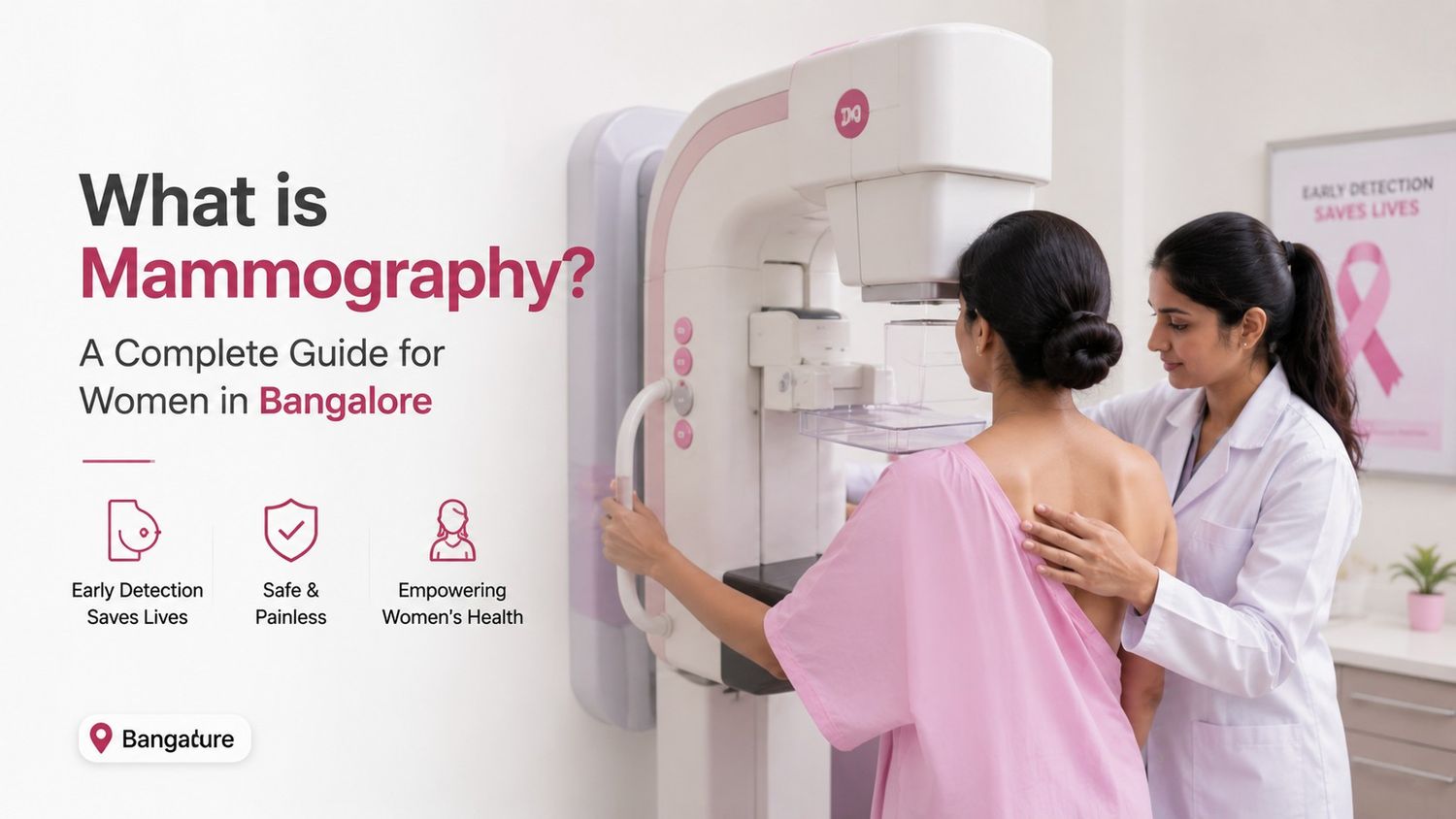
One of the most versatile and common test used in modern medicine is an X-ray test and now due to the advancements in the technology this technique is helping the doctors to analyze the patients without doing the surgery. Small amount of the radiations is used in X-rays to get the images of the body organs, bone and other internal structures. Thus the abnormalities can be easily determined with accuracy and more quickly with the help of this technique by the medical healthcare professionals. Besides being harmless X-rays are affordable, highly effective, fast and painless technique.  X-rays plays an important role in medical diagnosis and treatment planning such as in the identification of the fractures, in detecting lung infections etc.Following are the main reasons doctors recommend the X-ray tests:Fractures and Dislocations identification: One of the most common use of X-rays is the identification and dislocation of the fractures. Since the bones are denser than surrounding tissues, they absorb more radiation and stand out clearly on the image, and helps the doctors to determine the exact location of the break and severity of the fracture as well.Detecting Chest and Lung Conditions: For the patients experiencing the shortness of the breath, chest pain or chronic cough the chest X-ray is usually recommended by the doctors and this type of X-ray is vital for diagnosing the medical conditions like tuberculosis, heart failure, lung cancer and pneumonia etc.Joint issues monitoring and Diagnosis: In order to get an idea about the stiffness in the joints and pain in the knees, hips and hands etc.  the Joint X-rays are used to check the narrowing of joint spaces caused by arthritis or the gradual bone loss associated with osteoporosis. Swallowed Foreign objects locating in the body: An X-ray can even detect the swallowed metallic object if any by the patient it will help to quickly and accurately locate the item within the digestive or the respiratory tract of the patient so that the doctors can get an idea of the exact location and the treatment process can be started.Guiding of the surgical procedures: To ensure that broken bones are perfectly aligned or any medical implantation is rightly done in the areas such as artificial joints or the metallic plates in the correct position the X-rays aid drastically to the doctors in the same.Bone healing monitoring: With the help of the X-ray follow ups after the injuries or the surgeries the progress of the bone healing can be determined easily and this helps to ensure that the fusing of the bones at the right position is rightly done. Dental problems detection: Small scale X-rays are used by the dentists in order to determine the hidden tooth decay, cavities and to examine the roots of the teeth during the root canal process and it also helps to determine the health of the jawbone.Tumours and infections determination: Abnormal bone growths and masses within the body can be determined easily through the X-rays and any such infection and unwanted tumors in the body can be determined easily. Evaluating Heart Conditions: Heart conditions such as enlarged heart, fluid build-up, heart failure si8gns and abnormalities in the blood vessels can also be easily determined by the X-rays.Spine and Posture problems determination: Problems such as slipped discs, curvature of the spine, spinal fractures, spine diseases etc. can be easily determined though the X-rays and are widely used by the doctors.Determination of the Kidney stones and urinary problems: Problems such as kidney stones, bladder stones, blockages in the urinary tract can be determined easily through the X-rays.Chronic lung diseases like COPD are monitored easily by the X-rays.Safety of X-Ray TestsModern X-ray machines uses very low dose of the radiations, and thus the process of the X-rays is generally safe as compared to other techniques such as CT scan or an MRI scan. Only during the medical necessities, the X-rays are recommended by the doctors and protective measures such as lead aprons are often used to minimize exposure. Thus it is highly recommended by that any pre medical issue or history should be essentially told to the doctors before taking any X-rays especially during the pregnancy.ConclusionX-rays are widely recommended in the modern healthcare because of their wide applications right from diagnosing fractures and infections to detecting cancer and monitoring treatment progress, this technique provides the valuable information and insights to the medical healthcare professionals so that the effective treatment can be started immediately. X-ray procedure is quick, non-invasive and is highly effective tool used worldwide. They help in the better patient care and improved health outcomes and because of all these reasons X-rays are continuously recommended by the doctors for understanding and protecting human health.

Healthcare
Are X-Rays Safe? Benefits, Risks & Safety Tips Explaine...

One of the most common imaging technique used in the world is X-rays. They are widely used in the diagnosis of the diseases, injuries and helps in guiding the treatments accurately and quickly. X-rays play an essential role in modern healthcare right from checking broken bones to detecting infections in the lungs. Although the X-rays involve the exposure to radiations still it is safer and painless technique. Patients will feel more informed and confident when undergoing an X-ray examination if they are aware about the benefits, risks, and safety measures of the X-rays. X-rays are used to create the images of the internal structures of the body parts using the electromagnetic radiations. Lungs appear black on the X-ray images since they are spaces filled with air. To identify fractures, infections, tumours, dental problems, and other medical conditions these different absorption of the specific parts in the X-rays allows the doctors to use these contrasts in the early and easy diagnosis process.  It is non-invasive, fast and painless technique. It is one of the most efficient diagnostic tools available presently since the entire process takes only few minutes.Properties of X-Rays They have the same speed as that of the light.They cannot be deflected by electric or magnetic fields.Ability to ionize atoms and molecules.Can remove electrons from atoms.Potentially harmful if exposure is excessive.Benefits of X-RaysQuick and Accurate Diagnosis is possible with this technique.It is non-invasive and painless procedure.Helps in the detection of various conditions such as fractures, dental cavities, lung infections and breast cancer etc.Helps the doctors to access injuries after accidents.Helps in guiding the medical procedures.Helps to get the insides of the body’s internal structures.X-Rays are used to locate the swallowed objects etc.Are X-Rays Safe?When X-rays are used appropriately they are considered one of the safest and non-invasive technique. Thus ALARA principle (As Low As Reasonably Achievable) is used by the modern scanners to minimise radiation dose, particularly in children. The amount of radiation used in most routine X-rays is very small and unlikely to cause harm. Thus this imaging technique is comparatively safer and painless in comparison to other techniques such as CT scan or MRI.Risks of X-Rays Radiation Exposure: The amount of exposure is the biggest risk of the X-rays and if the unnecessary repeated scans are avoided the exposure risk can be minimized.Pregnancy Concerns: High radiation exposure during pregnancy may affect the developing baby, especially during early stages. Thus informing the doctors priory is recommended.Contrast Dye Reactions: Allergic reactions may be felt when the contrast dyes are used during the procedure and symptoms such as rashes, nausea and itching may be felt.Cumulative Effect: Higher radiation exposure over time get accumulated if the people undergo frequent imaging tests and thus maintaining the scans record is what is essentially recommended.Safety Tips for X-Rays Strict guidelines are followed by the medical professionals to ensure patient safety during X-ray procedures. Some of the safety tips for the procedure are:Using the Lowest Effective Dose.Protective Shields should be used during the scan.Limiting Exposure Time for the patient.Modern Digital Technology should be used for the imaging procedure.Careful Justification of the reports and other processes.Always inform your doctors if you are pregnant or have any other medical issues.It is highly recommended to keep the records of the previous X-rays for tracing the history of the patient in order to minimize the risk.ConclusionX-rays have transformed the medical healthcare by enabling the fast and accurate diagnosis and are most valuable and safer medical technique. They are widely used by the doctors to detect the infections, injuries and many other health conditions while guiding effective treatments. Modern technology and strict safety measures keep the risks very low since the very small amount of the radiations are used. Understanding the benefits and potential risks of X-rays allows patients to make informed healthcare decisions. Thus the safety measures should be used in order to avoid the risks and other potential myths of the X-rays and accurately medical information far outweigh the minimal risks associated with X-ray exposure.

Add Member
Provide email & phone to receive medical reports and notifications.