An MRI scan is a medical imaging technique that uses strong magnetic fields and radio waves to produce detailed images of the body’s internal structures. Soft tissues such as the brain, spinal cord, muscles, ligaments, and internal organs are usually examined by this technique. In the case of emergencies, MRI scan is widely used due to its detailed evaluation of the soft tissue injuries and CT scan is used because of the speed of the scan. An MRI machine is a large, tube-like structure with a flat, cushioned bed that slides in and out of the scanner. The bed moves in the machine slowly in which the patient lies.
A strong magnetic field and radiofrequency energy is used in the MRI scan instead of the ionizing radiations like that used in a CT scan or in an X-ray. MRI is used when the healthcare providers want to have a closer look at the soft tissues. Even the images of non-bony areas or soft tissues of the body can be easily taken by the MRI scans. It is best suited to examine the following areas:
· Brain
· Pelvis
· Spinal cord
· Nerves
· Ligaments and tendons
· Muscles
· Abdomen
An MRI is better than CT at distinguishing between different types of soft (non-bony) tissues. MRI scan images help the doctor to examine the detailed images of the tissues and structures inside your body. MRI scans are generally more expensive than CT scans or X-rays. Usually the MRI scans take longer time than the CT Scans.
Some common symptoms and medical conditions under which MRI scan is recommended to the patients are as follows:
- Numbness
- Weakness
- Injuries to head
- Headache
- Changes in the memory
- Joints pain
- Fractures
- Kidney stones
- Liver issues
- Fever due to unknown causes.
- Chest pain
- Blood clots
- Infections
Since the ionizing radiations are not used in the MRI scans healthcare providers often use these if a person requires frequent imaging to support a diagnosis and is particularly used for children or pregnant mothers.
Risks of the MRI Scan
Since a strong magnetic field is created in a MRI scan which can pose a safety risk if you have any medical history. You should always tell your healthcare provider if you have any metal in your body such as:
- Insulin pumps
- Pacemakers
- Cochlear implants
- Implanted cardiac defibrillator
- Deep brain stimulators
- Valgus nerve stimulators
Following are some of the things to be done post the MRI Scan are completed:
- If you were not sedated then the normal diet and activity can be resumed.
- If you took a sedative or had supervised sedation, do not drive, operate machinery.
- Keep the IV site clean and dry
- Drink water unless your clinician told you to restrict fluids.
CT is commonly known as Computed Tomography or CAT Scanning and it uses specialized X-ray technology to create detailed cross-sectional images of the body. These CT Scans can be considered as taking multiple X-ray slices that a computer assembles into comprehensive three-dimensional pictures of bones, organs, and soft tissues. These scans are quick at detecting tumours, identifying internal injuries, guiding treatment decisions in emergency situations and in diagnosing the vascular conditions. Since the X-rays are used in the CT scan more precision then the routine X-rays are used in this technique. CT scans are typically faster than MRI scans with the examination time in minutes only.
In order to get a closer look inside the head, skeletal system, and internal organs the healthcare providers use the CT Scans and to identify the following as well:
- Blood system disease
- Internal bleeding
- Infectious
- Inflammatory processes
- Injuries
Uses of CT Scans
CT scans are versatile in nature, and their uses include the following diseases diagnosis:
- Abscesses within different body areas
- Cancer screening and diagnosis
- Bladder and kidney stones
- Coronary artery diseases
- Blood clots
- Sinusitis
- Internal injuries from trauma
- Bone injuries
- Spine injuries
Safety Considerations and Risk Management
Following are the safety considerations during the CT scans:
· The principle of ALARA (As Low As Reasonably Achievable) guides protocols to minimize radiation while maintaining diagnostic quality.
· MRI safety centres on the magnetic field’s effects rather than radiation concerns
· The radiation exposure from CT Scans remains within the safe limits although it is higher than the regular X-rays.
· Always carry device identification cards and provide complete implant history.
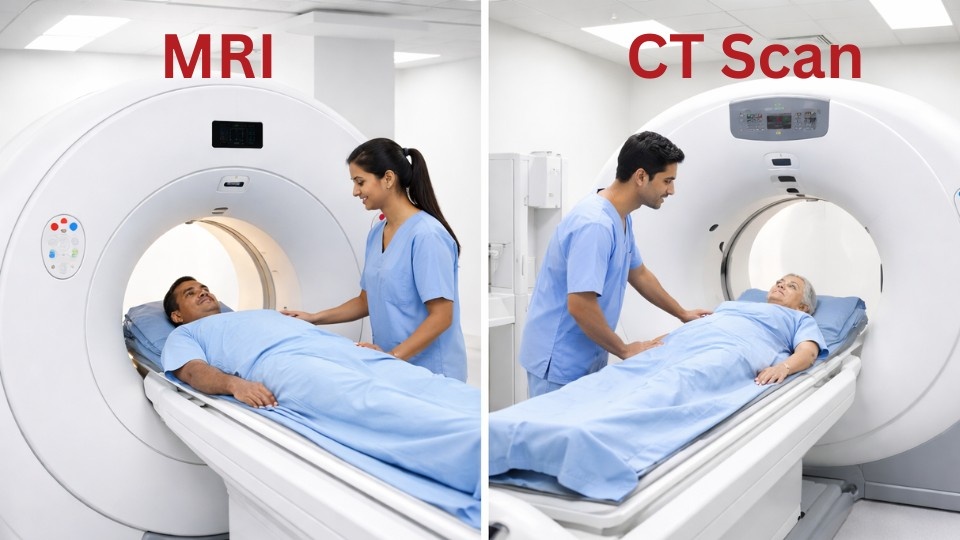
Main Difference between an MRI and CT scan
MRI :-
- Better for viewing of soft tissues, fat, water, and muscles.
- Takes more time for the images.
- More expensive.
- Uses strong Magnetic field.
- Can cause claustrophobia.
- Longer (typically 20 to 90 minutes.
CT Scan:-
- Optimal for viewing bones, such as the spine, and veins clot.
- Produce images faster.
- Less expensive.
- Uses ionizing radiations.
- Do not cause claustrophobia.
- Faster (typically 5 to 10 minutes).
Ionising radiation is used in CT Scanning. ALARA principle (As Low As Reasonably Achievable) is used by the modern scanners to minimise radiation dose, particularly in children. Usually the cumulative exposure is accounted in the CT scans because exposures from the CT scans are small. MRI uses a powerful magnet. Safety focuses on preventing metal movement or heating, protecting hearing, and avoiding burns from skin-to-skin or cable loops. All patients complete a detailed MRI safety checklist.
Choosing between an MRI and a CT scan
Based on your symptoms the doctor will most likely recommend whether you should get an MRI or a CT scan and when the detailed images of your ligaments, organs and soft tissues are needed the doctor is likely to suggest an MRI scan to you and it includes the cases such as torn ligaments, soft tissues and herniated disks while a CT scan will commonly be recommended if the images of the internal organs, or due to a fracture or head trauma is needed by the doctor generally.