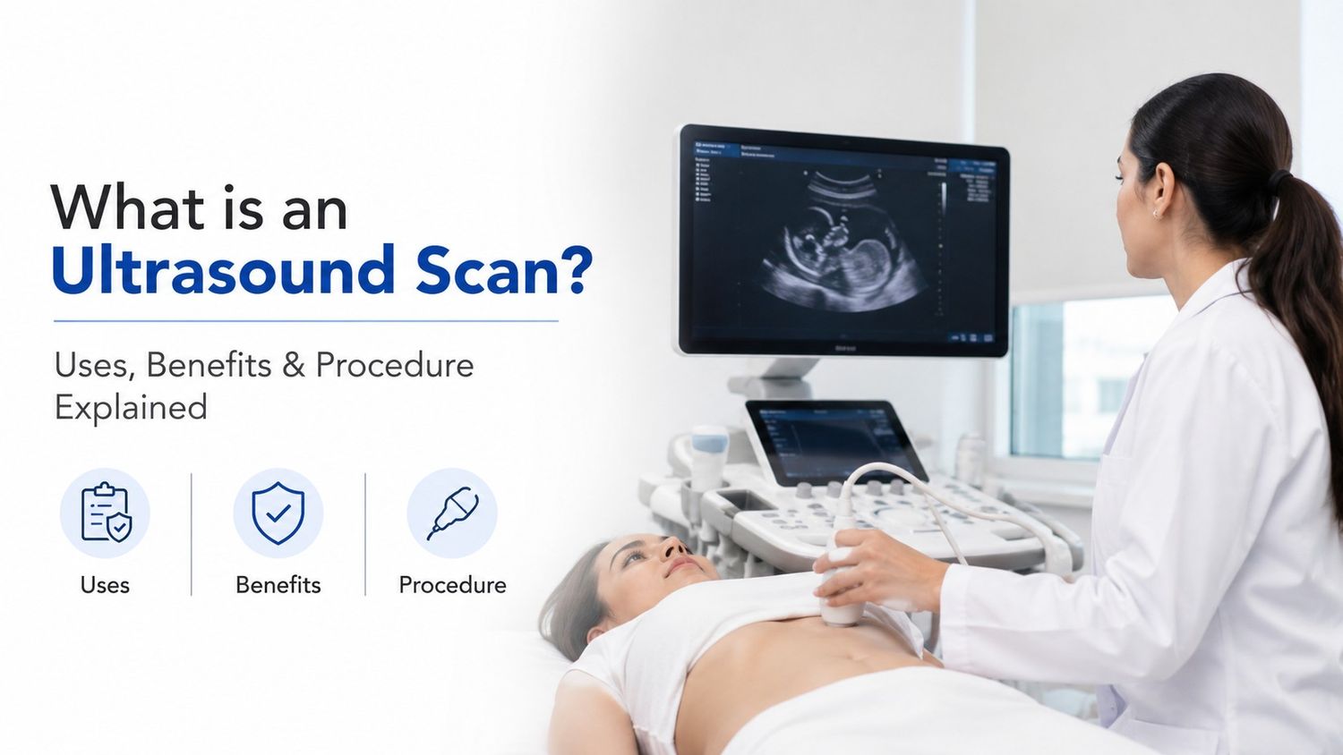
An ultrasound is the painless imaging test used to examine internal organs and tissues of the body it is also known as Sonography or Ultrasonography. This technique uses high frequency sound waves to create the real images of the internal structures of the body. It is one of the safest diagnostic techniques available today since it does not uses harmful ultraviolet radiations. To diagnose medical conditions, monitor pregnancy, guide certain medical procedures, and examine body organs such as the liver, kidneys, heart, and reproductive organs these scans are widely used in clinics, diagnostic centres and in the hospitals. In some cases the ultrasound is taken by inserting certain devices inside the body.
Procedure of the Ultrasound
A device called transducer is placed on the skin during the ultrasound scan. The high frequency sound waves are sent by the transducer into the body. These sound waves bounce back after hitting organs and tissues. The images are displayed by the computer on the screen when it receives the echoes from the device and then converts them accordingly. The shape, size, and condition of internal organs can be easily observed by the doctors during the scan since each tissue reflects sound waves differently.
Types of Ultrasound Scans
- Abdominal Ultrasound: To examine the internal organs like liver, spleen and kidneys.
- Pelvic Ultrasound: To study the reproductive organs like uterus and ovaries in women and prostate gland in men.
- Pregnancy Ultrasound: To monitor the growth and development of the baby.
- Doppler Ultrasound: To check the circulation problems and measure the blood flow in the blood vessels.
- Echocardiography: A special ultrasound used to examine the heart and its functioning.
- Thyroid Ultrasound: To detect thyroid nodules, swelling, or abnormalities.
Procedure of an Ultrasound Scan
The procedure of the ultrasound depends on the type of the body part under examination but the overall procedure remains the same:
- Step 1: Preparation for the scan.
- Step 2: Application of Gel on the area under examination.
- Step 3: Use of Transducer on the skin in order to start the scanning procedure.
- Step 4: Image Formation by the computer on the screen using the echoes of the scans.
- Step 5: Completion of Scan and obtaining the results for analysis.
Preparation before an Ultrasound Scan
Preparation depends on the type of ultrasound being done but the general guidelines remains the same:
- Avoid eating for 6–8 hours before the abdominal scan.
- Drink plenty of water to keep the bladder full if you are undergoing a pelvic ultrasound.
- A full bladder may be required in early pregnancy in order to undergo the ultrasound scan during pregnancy.
- Avoid smoking or caffeine before the Doppler ultrasound as they affect blood flow.
Post ultrasound
- Review of the images by the doctor.
- Appointment for the results discussion and to discuss the new findings if any in the results.
- If other diagnosis needed or not such as a CT scan, MRI, or a biopsy sample of tissue depending on the area examined.
- Beginning of the treatment immediately by the doctor.
Benefits of Ultrasound Scan
Ultrasound scanning offers several advantages which are as follows:
- Radiation-Free imaging technique.
- Painless and Non-Invasive diagnosis tool.
- Real-Time Imaging.
- Safe during Pregnancy.
- Quick and Convenient diagnostic tool.
- Cost-Effective.
- Helps in Early Diagnosis.
- Portable Technology.
Uses of Ultrasound Scan
Following are the main uses of the Ultrasound scan:
- Confirms pregnancy
- Monitoring of the pregnancy
- Monitors baby growth
- Detects abnormalities
- Checks amniotic fluid levels
- Liver diseases
- Kidney stones
- Gallstones
- Pancreatic disorders
- Spleen abnormalities
- Detecting Tumours and Cysts
- Identify abnormal growths in organs
- Check the Blood clots
- Blocked arteries are identified
- Echocardiography helps to access the problems of heart valve
- Congenital heart diseases
Limitations of Ultrasound Scan
Although ultrasound is highly useful, it has certain limitations which are as follows:
- Behind the bones cannot be clearly imaged by using the ultrasound scan.
- For obese patients it is less effective.
- Air-filled organs like lungs are difficult to examine.
- Sometimes additional tests like CT or MRI may still be needed for the further treatment.
Conclusion
One of the most important and widely used diagnostic tools in modern medicine is the Ultrasound scanning. It is safe, painless, affordable, and highly effective for examining internal organs, monitoring pregnancy, and diagnosing various medical conditions. Ultrasound plays a vital role in healthcare from detecting organ abnormalities to assessing blood flow and guiding medical procedures. With advancements in technology, ultrasound machines now provide clearer images and faster results, helping doctors diagnose diseases accurately and begin treatment at the right time.