The technique in which the high-frequency sound waves are used to create images of structures inside the body is known as sonography or Ultrasound and it’s the widely used medical imaging technique. Ionizing radiations unlike in the CT Scans and X-Rays are not used in the ultrasound and thus this technique is safe, painless and non-invasive diagnostic tool and thus technique helps in the early detection of the diseases and helps in the monitoring of the internal organs and helps in guiding the medical procedures. The principle used in an Ultrasound is echoes and a device called transducer is used to emit the high-frequency sound waves into the body. These sound waves travel through the tissues and are reflected back when they hit boundaries between different tissues the returning echoes are recorded and captured and in turn they convert the real time images on a monitor and the image for the same is obtained through the ultrasound. It is commonly used in the clinics, hospitals and diagnostic centres because of its ability to provide the instant results and is safer technique to use.
Types of Ultrasound
Based on its application and technique ultrasound can be classified as follows:
- Diagnostic Ultrasound: This is the ultrasound used to visualise the internal organs such as liver, kidneys and uterus and is the most common type of the ultrasound. This type of the ultrasound can give the 2D, 3D and 4D images easily depending upon the organ under examination. 3D ultrasound is used to observe fetal structures during pregnancy while the 4D ultrasound is used to see the movement of the baby in the Mother’s womb.
- Doppler Ultrasound: This type of ultrasound is used to measure the movement of the blood through the blood vessels. In this colour is used and blood flow, direction and speed is shown by different colours. This type of ultrasound is used to detect low blood flow and is more sensitive in nature. Blockages, clots, and heart conditions are especially detected by this type of ultrasound.
- Therapeutic Ultrasound: This type of ultrasound is used in physiotherapy, for reducing the pain and inflammation and for breaking the kidney stones as well.
- Specialized Type of Ultrasounds: Other than these types of ultrasounds there are more specialised ultrasound types which are used in special cases such as:
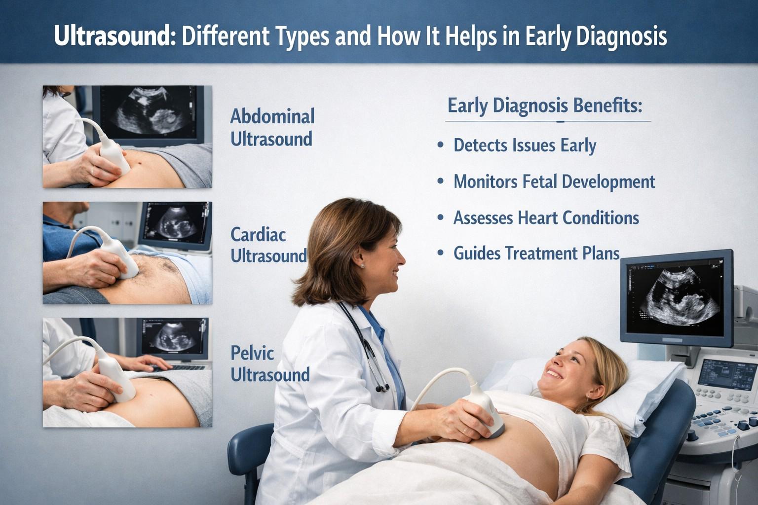
- Echocardiography: It is used to detect abnormalities in heart structure and function.
- Obstetric Ultrasound: Used to monitor fetal development during pregnancy.
- Abdominal Ultrasound: Organs like the liver, pancreas, and kidneys are examined through this type of ultrasound.
- Transvaginal Ultrasound: The detailed images of female reproductive organs are provided by this type of ultrasound.
Body parts where the Ultrasound is widely used by the doctors for Diagnostics
Ultrasound is used by the Physicians to help diagnose conditions affecting the organs and soft tissues of the body, including:
- Gallbladder
- Thyroid
- Pancreas
- Breast
- Bladder
- Ovaries
- Eyes
- Testicles
- Liver
- Abdomen
- Heart
- Spleen
- Kidneys
- Blood vessels
Besides lot of uses of the ultrasound it has certain limitations as well because sound waves are not able to transmit the through areas that may hold gas or air (like intestines), or areas blocked by dense bone.
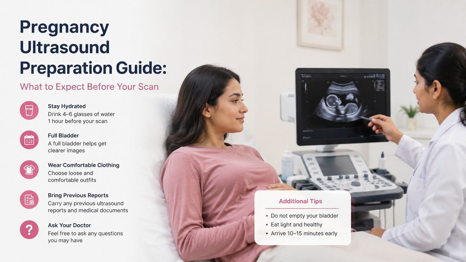
Preparation for the Ultrasound
Depending on the area or the organ being examined the preparation of the sonography is dependent and based on these organs the different preparations are used and carried out:
- Fasting for 8-12 hours before the ultrasound is conducted can be advised by the doctor.
- Consumption of the fat tree meal and fast till the procedure is required for the liver and pancreas diagnosis.
- Water intake and extreme medication might be advised to the patients.
- For viewing the pelvic organs the individual might be asked to drink lot of water.
Procedure used for the Ultrasound
Following steps are used for the Sonography which is as follows:
- Specialised dresses are used for the ultrasound so that patient feels the comfort.
- In order to prevent the friction and rub the ultrasound transducer on the skin a specialised lubricating jelly is used which helps in the transmission of the sound waves.
- The reverberations are reflected into a computer.
- Picture is formed by the sound waves on the monitor and high resolution images for the same are used.
- Depending on the area being examined, the change in the position might be needed in order to get the better access of the area under examination.
- In about 30 minutes the gel is cleared off from the skin after completing the ultrasound process.
Procedure used after the Ultrasound
The images are checked and reviewed after the ultrasound is carried out for any abnormalities by the doctor. If any abnormalities seen in the ultrasound the help of other techniques such as MRI, CT scan, or a biopsy sample of tissue depending on the area under examination can be used in order to get the better results for quick and early diagnosis.
Applications of Using the Ultrasound for the Early Diagnosis
- Abnormal Growth and its detection such as tumours, cysts etc.
- Diagnosis of blockages and blood clots.
- Monitoring Pregnancy and foetal growth and development.
- Early diagnosis can prevent serious conditions like heart attacks or strokes.
- To identify the disorders of the organs.
- Infections and Inflammation detection.
- Medical Procedures guidance.
- Screening and Preventive Healthcare.
Advantages of Ultrasound in Early Diagnosis
- It is safe since it uses no harmful radiations.
- Immediate results are obtained.
- No surgery is required and thus it is non-invasive in nature.
- It is cost effective technique and easily affordable.
- It is portable in nature and can easily be used in emergency.
Limitations of Ultrasound
Besides being useful it has some limitations as well:
- Operator’s skill determines the image quality.
- Very small or deep abnormalities may not be detected easily and avoided.
- Behind the bones and air -filled organs the image structures cannot be seen clearly.
Even after so many limitations this technique remains one of the most widely used diagnostic tools because of its non-invasive, safe and cost effective nature. It is most accessible method for the human body imaging. Thus it is quite important for diagnosing serious diseases like tumours and heart conditions, such as blockages, blood clots to detect the presence of cysts and tumours. Thus it is a versatile technology and it is of utmost importance in early diagnosis and medical care.