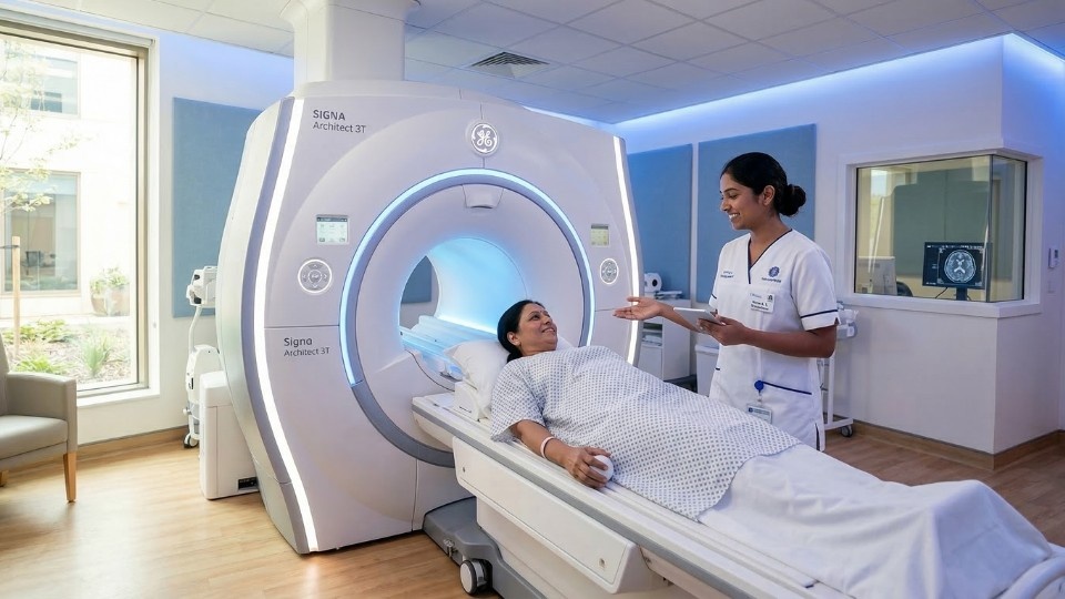
In order to determine what your body is undergoing without undergoing surgery is aided by MRI and CT scans they help to monitor the progress of the body diagnosis towards a particular treatment and making the right choices for the same. Anxiety and stress are the common symptoms when entering for the scans and knowing the consequences of the scans before, during and after the scan is very important for the individual point of view so that the patient can feel relax and calm so that anxiety and other harmful symptoms can be reduced. The technique which uses computer technology and X rays so as to create the detailed cross-sectional images of the body and it is a medical imaging technique used in the present time. It helps doctors examine bones, organs, and blood vessels and detect conditions such as fractures, tumours, internal bleeding, or infections. An MRI scan is a medical imaging technique that uses strong magnetic fields and radio waves to produce detailed images of the body’s internal structures. Soft tissues such as the brain, spinal cord, muscles, ligaments, and internal organs are usually examined by this technique. In the case of emergencies, MRI scan is widely used due to its detailed evaluation of the soft tissue injuries and CT scan is used because of the speed of the scan. An MRI machine is a large, tube-like structure with a flat, cushioned bed that slides in and out of the scanner. The bed moves in the machine slowly in which the patient lies.
Understanding the MRI Scans
For the better understanding of the condition of the patient doctor usually recommends the MRI scan to the patients because the MRI scan images helps the doctor to examine the detailed images of the tissues and structures inside your body. Therefore the interpretation of the MRI scans results is very vital and is supportive for the doctor in order to make a decision for the accurate diagnosis. The cases where the symptoms shows the condition is related to nerves, joints, or internal organs in such cases the MRI scans aid drastically since it produces the images with high resolution and clarity.
There are some common symptoms and medical conditions under which the MRI & CT scan is recommended to the patients are:
- Changes in the memory, injuries to the head, headache.
- Weakness and numbness.
- Fractures and joint pains including the common sports injuries.
- Fever due to unknown causes.
- Infections, chest pain and blood clots.
- Kidney stones, common liver issues.
- Cancer detection, staging, and treatment follow-up.
Ionising radiation is used in CT Scanning. ALARA principle (As Low As Reasonably Achievable) is used by the modern scanners to minimise radiation dose, particularly in children. Usually the cumulative exposure is accounted in the CT scans because exposures from the CT scans are small. MRI uses a powerful magnet. Safety focuses on preventing metal movement or heating, protecting hearing, and avoiding burns from skin-to-skin or cable loops. All patients complete a detailed MRI safety checklist.
The precautions usually undertaken by the doctor for these scans are as follows:
- Kidney disease: MRI and CT scans impact the patients suffering from kidney disorders.
- Implants/devices: Some pacemakers, , or older implants are unsafe for the MRI whereas the modern MRI devices are safe along with these implants as well and they interfere less.
- Pregnancy: MRI and CT scans are generally avoided during the pregnancy since CT involves radiation ,unless absolutely necessary
- Patches with metal foils, tattoos, piercing and some cosmetics interfere with the MRI scanning and to avoid such situations the screening is essential before undergoing such scans.
In order to ease the anxiety before such scans and from the emotional point of view of the patient and it should be used under the controlled process so as to ease the anxiety before, during these scans:
- Slow diaphragmatic breathing.
- An eye mask can also be used to see inside the scanner.
- A blanket or cushion can be used for the comfort during the scan.
- Oral anti-anxiety medication or supervised sedation in advance can also be discussed.
CT Scan Process involves the scanning, administration and positioning of the images and for the patient as well it is very common to feel uncomfortable during the scans and at times you may feel the tiredness and in such case immediately contact the doctor if you have:
- Hives, swelling of lips/tongue, trouble breathing, chest tightness.
- Severe or worsening headache, confusion, or fainting.
- Persistent nausea/vomiting, fever, or chill.
- Painful swelling, redness, or skin blistering at the IV site.
- Little or no urine output for 24 hours after contrast, especially if you have kidney disease.
For all the things to go smoothly the right outfit is must and it reduces the chances to change as well. In such cases avoid wearing watches, jewellery or the metal accessories because the metal objects interfere with the MRI machine. For the interpretation of the reports results an advice is always recommended because the interpretation of the results is not easy.
Based on the report, your clinician may:
- Refer you to a specialist (e.g., neurology, orthopaedics, and cardiology).
- Start physical therapy or change the lifestyle approaches.
- Recommend surgery or image‑guided procedures.
- Schedule follow‑up imaging to monitor changes.
- Adjust medications as per the undergoing processes and treatment under process.
In order to deeply analyse the results to determine the in depth analysis of the root cause of the particular disorder repeated scans can also be done.
Following measures should be taken in such cases:
- Keep a record of past imaging, reports, and CDs/USBs; bring them to new appointments.
- Share your implant cards and exact device models.
- Tell staff about all allergies, kidney issues, medications, pregnancy status, and tattoos/patches.
- Ask if non‑contrast imaging or alternative tests could answer the question.
Alternatives to Consider: Ultrasound, X-ray, or Delayed Imaging Based on the situation of the patient can be considered as the alternatives for the MRI Scan. Following are some of the potential alternatives:
- Ultrasound: No radiation, good for gallbladder, pregnancy, blood flow (DVT), thyroid, and some soft‑tissue lumps.
- X-ray: Quick for bones, chest, and device checks; less detail than CT/MRI.
- Non‑contrast protocols: May answer many questions when contrast is risky.
Following are some of the things to be done post the MRI Scan is completed:
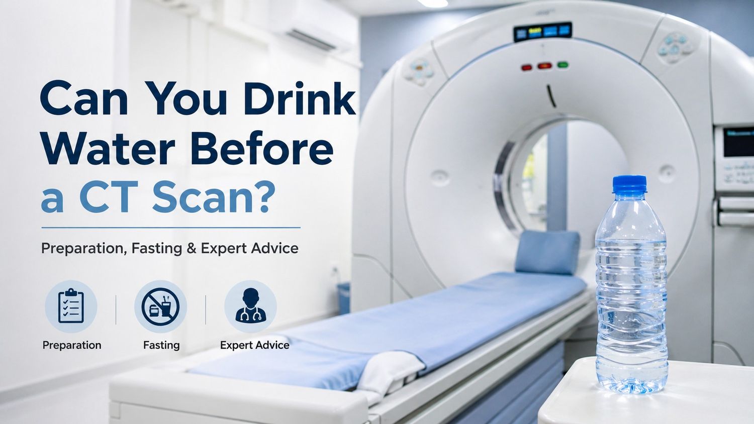
- Drink water unless your clinician told you to restrict fluids.
- Resume normal diet and activity if you were not sedated.
- If you took a sedative or had supervised sedation, do not drive, operate machinery.
- Keep the IV site clean and dry; mild soreness is common.
- Seek prompt care for hives, swelling, breathing trouble, chest pain, severe headache, fever, worsening redness or very low urine output.
The results are usually obtained in a week. The MRI images is usually reviewed by a radiologist, who is a board certified specialist trained in interpreting medical images. This process usually takes several days, as the radiologist will carefully examine each image, and compile a report to share with your healthcare provider or the doctor.
Understanding the CT Scans
CT is commonly known as Computed Tomography or CAT Scanning and it uses specialized X-ray technology to create detailed cross-sectional images of the body. These CT Scans can be considered as taking multiple X-ray slices that a computer assembles into comprehensive three-dimensional pictures of bones, organs, and soft tissues. These scans are quick at detecting tumours, identifying internal injuries, guiding treatment decisions in emergency situations and in diagnosing the vascular conditions.
For the older people CT scans frequently diagnose conditions such as pulmonary embolism, kidney stones, abdominal pain causes, and bone fractures that might not appear clearly on regular X-rays. It is usually preferred for the elders because of its speed of scanning in comparison for the X-rays which makes it easy for them to undergo these scans during the treatment process and also easily feasible for those experiencing acute symptoms requiring rapid diagnosis. Brain CT scans can quickly identify strokes, bleeding, or tumours, while chest CTs provide detailed lung imaging crucial for diagnosing respiratory conditions common in older adults.
Safety Considerations and Risk Management
Following are the safety considerations during the CT & MRI scans:
· The radiation exposure from CT Scans remains within the safe limits although it is higher than the regular X-rays.
· The principle of ALARA (As Low As Reasonably Achievable) guides protocols to minimize radiation while maintaining diagnostic quality.
· MRI safety centres on the magnetic field’s effects rather than radiation concerns
· Always carry device identification cards and provide complete implant history.
Lot of Myths about MRI and CT scan are there, some of them are as follows:
Myth: MRI uses radiation like a CT.
Fact: MRI uses a magnetic field and radio waves—no ionizing radiation.
Myth: All pacemakers make MRI impossible.
Fact: Many modern devices are MRI‑conditional with proper protocols.
Myth: CT contrast always harms the kidneys.
Fact: For most people with normal kidney function, risk is very low.
Myth: Claustrophobia means I can’t have an MRI.
Fact: Comfort techniques, wide‑bore scanners, and short‑acting medication can make MRI possible for most people.
For managing the senior health conditions powerful diagnostic tools such as MRI or CT scan are used which provides crucial information. Understanding these scans usually helps to understand what to expect in these scans and clear communication with your doctor helps greatly in this. Ultimately, the brief discomfort or anxiety associated with CT or MRI scanning pales in comparison to the valuable diagnostic information these tools provide. The quality of life and improved treatment outcomes helps in the early detection and accurate diagnosis. Thus these technologies help to maintain health and solve the difficulties in the body functioning as early as possible.