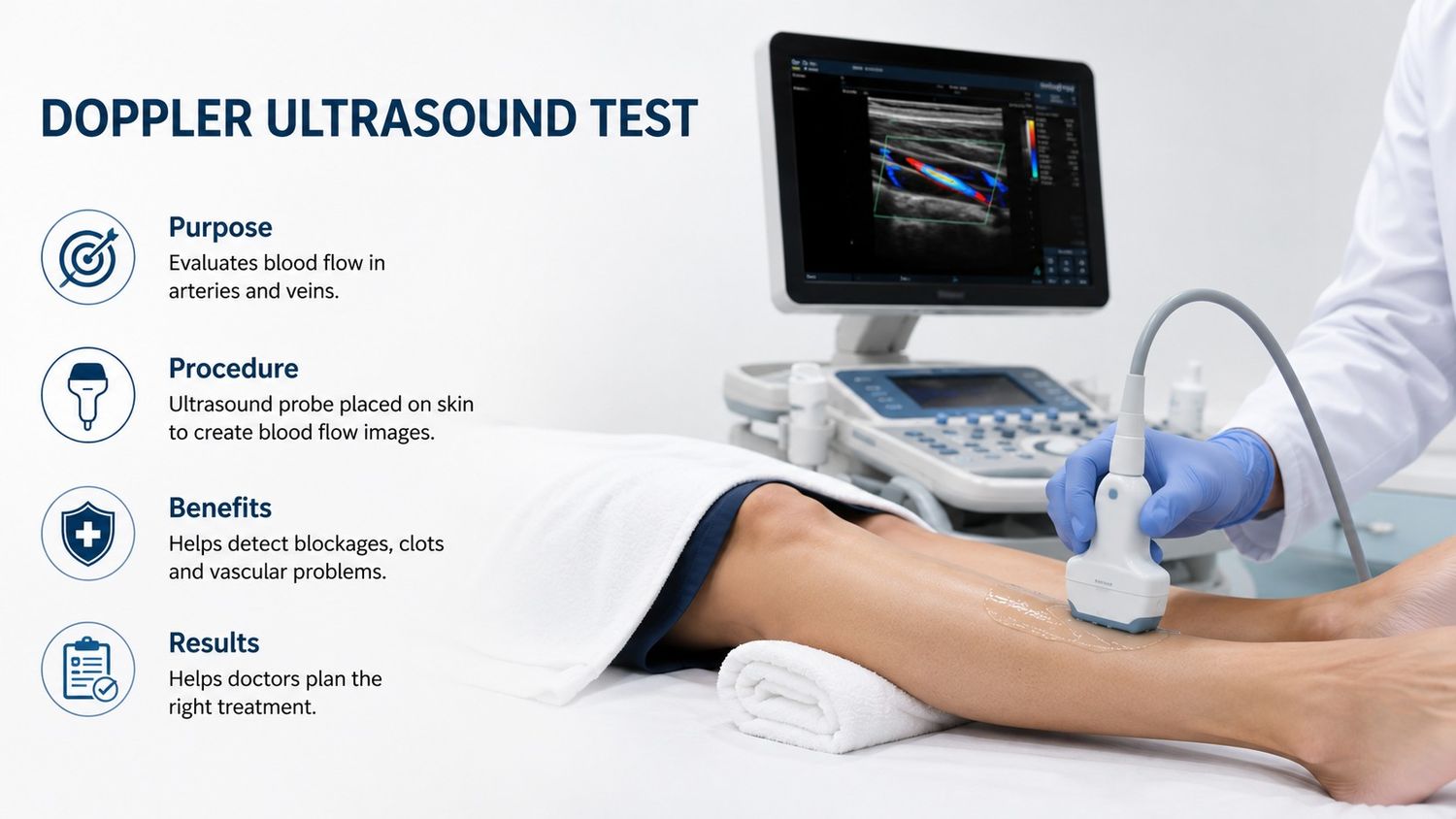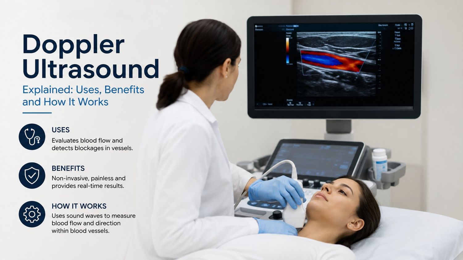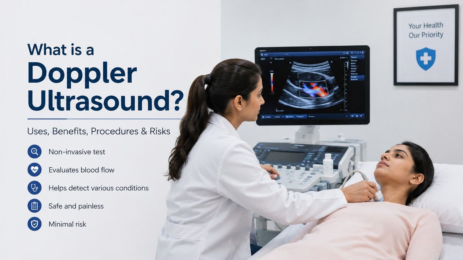
Doppler Ultrasound is the non-invasive technique which uses high-frequency sound waves to evaluate blood flow through the body's blood vessels. The Doppler ultrasound measures the movement of the blood within arteries and veins unlike the standard ultrasound which creates the images of the tissues and the organs. It is widely used in the diagnosis conditions related to pregnancy, heart issues and blood vessels. Diagnosis of various vascular and cardiac conditions can be determined easily through this ultrasound as it helps to determine the abnormalities in blood circulation.
It is widely used in the hospital and diagnostic centers to assess the blood flow in the arteries, determine the blockages and monitor the pregnancy and helps to evaluate the heart health.
Preparation for a Doppler Ultrasound
The preparation for the tests includes the following requirements such as:
- Loose clothing is usually recommended for the patients for smooth processing.
- Fasting is usually recommended by the doctor for patients if it involves the abdominal blood vessels.
- For any medications if any please inform your doctor in advance.
- Staying hydrated is recommended during the ultrasound.
- Avoid applying the lotions on the area being scanned.
- Provide the medical information to your doctor before starting the procedure.
Risks involved: Although it is the safest technique and is painless as well. Till date there are no known harmful effects associated with the procedure when performed by trained professionals. No discomfort is seen in the patients except the slight pressure from the transducer. Thus there are no major side effects of the ultrasound and in most of the cases the normal activities can be resumed immediately after the test.
Advantages of Doppler USG Scans
Doppler ultrasound has the following advantages such as:
- Non-Invasive technique.
- Radiation-Free procedure and is safe.
- Real-Time Results are obtained easily.
- Versatile technique
- Cost-Effective in nature.
- Early Detection of the abnormalities is easy.
- Guidance for Treatment is provided through the assessments.
Types of Doppler Ultrasound
- Spectral Doppler Ultrasound: It displays the flow of blood in the body in the form of the graphs and thus allows the doctors to measure the velocity of the blood flow.
- Colour Doppler Ultrasound: The direction and the speed of the blood flow is determined using the colour coding.
- Power Doppler: It is more sensitive than the colour Doppler and it can detect the slower blood flow as well.
- Duplex Doppler Ultrasound: It makes the use of the traditional ultrasound technology with the Doppler technology.
- Continuous Wave Doppler: It is quite useful for measuring high-speed blood flow and it continuously sends and receives the sound waves.
General Procedure for Doppler Ultrasound
The general procedure for Doppler Ultrasound Scans are as follows:
- The patient is asked to lie down on the examination table before the ultrasound.
- The probe is placed by the radiologist on the concerned area.
- The blood flow is measured by the pitch of the sound.
- Graphs and pictures are created on the screen by the machine.
- High frequency sound waves are used by the radiologists.
- The radiologist may ask you to change positions during the procedure to get clearer images from different angles for the specific areas.
- The entire procedure takes around 20-45 minutes.
- The gel is wiped out of the skin once the scan is completed.
Under what conditions can Doppler ultrasound help diagnosis
Doctors recommend the Doppler ultrasound to diagnose:
- Narrowed arteries or veins.
- Blood clots, including deep vein thrombosis (DVT).
- Blood vessel injuries.
- Chronic venous insufficiency (CVI).
- Evaluate blood supply to a transplant organ like kidneys and pancreas.
- Tumours in blood vessels.
Conclusion
Thus it can be concluded that Doppler Ultrasound has wide applications and benefits in the field of medical science and is one of the most useful and non-invasive technique since the exposure to radiations is not there it is completely safe for the pregnant women, children and old age people as well. The extensive applications of this technique makes it so useful along with its cost effective advantage and the interpretation of the results is so easy which further helps in the diagnosis of the treatment after the detection of the abnormalities.