A medical test that uses high high-frequency sound waves to capture live images from the inside of a patient’s body is known an Ultrasound and is also known as sonography. This imaging technique is similar to the technique of the radar and solar used by the military to detect planes and ships. It helps the doctors to see problems inside the body of the patient without making an incision such as the problems with vessels, tissues and organs. No radiations are used in this technique unlike the other techniques such as CT scan or X-rays and thus it is a preferred method for viewing a developing foetus during pregnancy since it is the safe technique.
Why an ultrasound is recommended?
If the patient is having pain, swelling, or other symptoms that require an internal view of the patient’s organs an ultrasound is recommended by the doctor. In case of the pregnant women it helps the mother to get the first view of her unborn child.
An ultrasound is used for the following:
- Bladder
- Eyes
- Gallbladder
- Kidneys
- Liver
- Ovaries
- Pancreas
- Spleen
- Thyroid
- Testicles
- Uterus
- Blood vessels
- In infants its brain
Procedure for an ultrasound
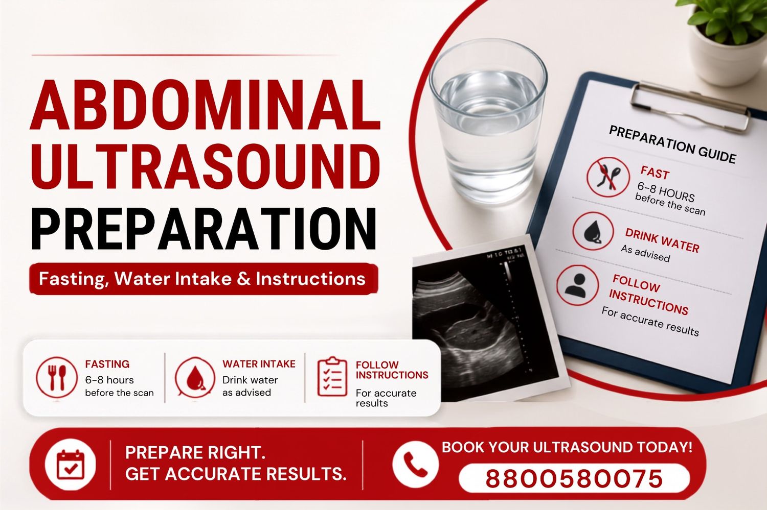
- Preparation before the Test: Wear the loose and comfortable clothes.
- Entering the Ultrasound Room: In the comfortable position the patient is allowed to lie down before starting the scan.
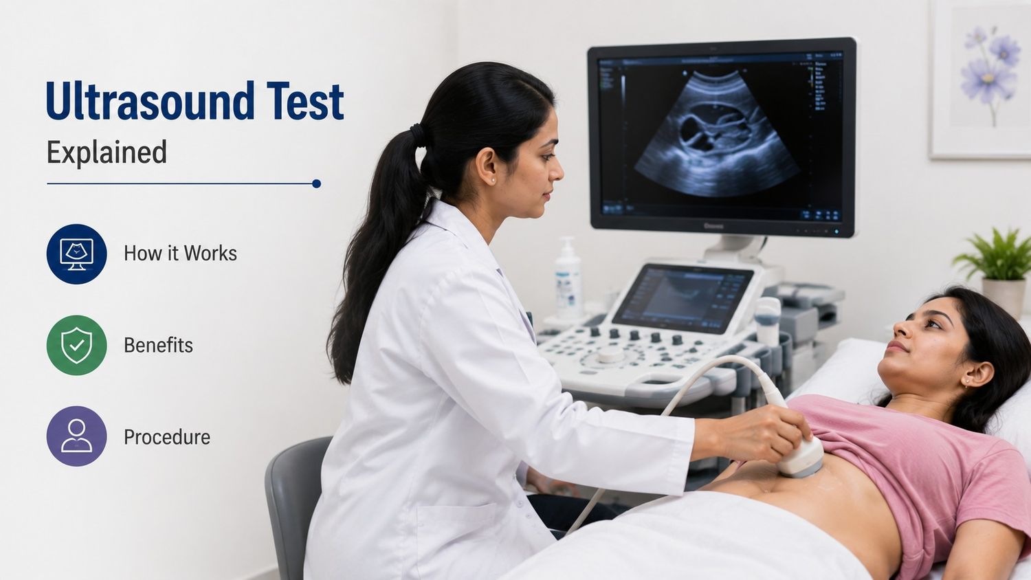
- Application of Gel: A gel is applied on the area which is to be scanned in order to get the better images and it helps the sound waves to travel properly.
- Use of the Transducer: A transducer is moved over the gel-covered area. The transducer sends and receives sound waves.
- Formation of Images: The images of the scanned portions are formed on the computer screen by using the echoes which are bounced back by the tissues and organs.
- Observation and Examination: The structures, movement and the exact condition of the organs is observed and examined by the technicians.
- Completion of the Procedure: The entire scanning process completes in about 15-45 minutes and is largely based on the type of the scan being done.
- Results and Diagnosis: The recorded images are examined by a radiologist or doctor, who prepares a report to help diagnose medical conditions or monitor treatment progress.
How ultrasound works?
- High-frequency sound waves are used in the Ultrasound.
- A special device called a transducer is used to get the improved images.
- The transducer sends sound waves to the monitor.
- When the sound waves hit different body parts of the body the echoes are produced.
- The transducer receives these echoes from these body parts under examination.
- The computer converts the echoes into images.
- This procedure is painless and safer technique.
Uses of Ultrasound Scan
Ultrasound scans are used for diagnosing and monitoring many health conditions such as:
- Determines baby position during the pregnancy.
- Examines the poor circulation of blood during the circulation process if any.
- Heart pumping function is also examined by the ultrasound scan.
- Ultrasound is often used during fluid drainage.
- Detects the abnormalities during the initial days of pregnancy.
- Checks amniotic fluid levels during pregnancy.
- Use to examine the organ disorders such as gallstones and kidney stones.
- Spleen abnormalities are also detected by the ultrasound.
- Ultrasound helps identify lumps, cysts, or abnormal growths in organs.
- Blood clots and arteries are detected through an ultrasound.
- Echocardiography helps to access congenital heart diseases.
- Ultrasound is often used for guiding the medical procedures like injections.
Difference between Ultrasound and Other Imaging Tests
- Uses sound waves
- No radiation
- Safe in pregnancy
- Affordable
- Real-time imaging
- Uses X-rays
- Radiation exposure is present
- Usually avoided in pregnancy
- Costlier
- Detailed body imaging
- Uses magnetic fields
- No radiations are there
- Sometimes restricted
- More expensive
- Excellent soft tissue imaging
Conclusion
Thus we can conclude that ultrasound is the painless imaging test used to examine internal organs and tissues of the body and is the most important tools used in the modern medicine. It is safer, painless and no radiation technique. It helps to determine the abnormalities which are not detected easily by other imaging techniques and is most affordable techniques. Ultrasound machines now provide clearer images and faster results because of the growing technological changes. These scans are widely used in clinics, diagnostic centres and in the hospitals and in some cases the ultrasound is taken by inserting certain devices inside the body.