Health packages
Add Member
Provide email & phone to receive medical reports and notifications.

Book your Appointment!
Icon Select Test
Icon
Not sure what you need?
+91 88005 80075
Let us know your health conditions. Our health advisor guide you.
Back SERVICES

VER/VEP

The VER/VEP test is a simple yet powerful tool for detecting problems in the visual pathways. By recording how your brain responds to visual signals, it helps doctors identify eye and neurological conditions early often before symptoms become severe.

Visual Pathway Assessment
Detects Eye & Brain Disorders
Non-Invasive & Safe
Expert Neurologist Report
VER/VEP

VER/VEP Test: Understanding Visual Evoked Potentials

Have you ever wondered how doctors can measure how well your brain responds to what your eyes see? That’s exactly what a VER/VEP test does.

VER (Visual Evoked Response) or VEP (Visual Evoked Potential) is a simple, non-invasive test used to check the visual pathways between your eyes and brain. It plays a crucial role in diagnosing and monitoring eye and neurological conditions.

What is a VER/VEP Test? +

The VER/VEP test measures the brain’s electrical activity in response to visual stimuli, such as flashing lights or patterned images.

  • Visual Evoked Potential (VEP): The electrical signals recorded from the brain when the eyes are stimulated by visual input.
  • Visual Evoked Response (VER): Another term often used interchangeably with VEP.

This test helps determine if there are problems with the optic nerves or visual pathways that carry signals from your eyes to your brain.

Why is a VER/VEP Test Done? +

Doctors may recommend a VER/VEP test to:

  • Diagnose optic neuritis (inflammation of the optic nerve)
  • Detect multiple sclerosis (MS) in early stages
  • Evaluate unexplained vision loss
  • Check for optic nerve damage after trauma
  • Assess visual development in children
  • Monitor glaucoma or other eye conditions affecting nerve function

πŸ“Š Fact: Studies show that VEP testing can detect optic nerve dysfunction even before symptoms of vision loss appear.

How Does a VER/VEP Test Work? +

The Process

Preparation

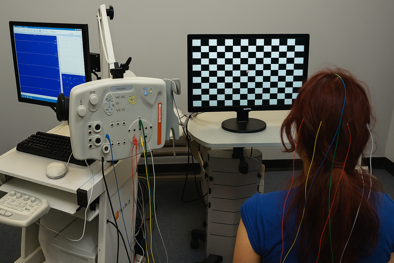
Electrodes are placed on your scalp (similar to an EEG).

You’ll sit in front of a screen displaying checkerboard patterns or flashing lights.

Stimulation

The patterns or flashes stimulate your visual system.

The electrodes record how quickly and effectively your brain responds.

Analysis

The recorded signals are analyzed to see if they fall within normal response times.

Delayed or weak responses may indicate nerve damage or disease.

What to Expect During the Test? +
  • Duration: Usually 30–45 minutes
  • Discomfort: Noneβ€”it's painless and non-invasive
  • Preparation: No special preparation needed, but avoid heavy makeup or hair oils (as electrodes need to stick to the skin)
  • Safety: Completely safe, with no side effects

πŸ’‘ Tip: Bring your glasses or contact lenses if you use them, since clear vision ensures accurate results.

What are the Benefits of VER/VEP Testing? +
  • Early detection of optic nerve and brain-related disorders
  • Non-invasive and safe with no radiation exposure
  • Helpful for children and patients unable to communicate vision problems
  • Supports treatment decisions by tracking disease progression
What are the Limitations of VER/VEP? +
  • Cannot pinpoint the exact location of the problem
  • May be affected by poor vision due to cataracts or uncorrected refractive errors
  • Best used in combination with other tests (MRI, eye exams) for accurate diagnosis
Add Member
Provide email & phone to receive medical reports and notifications.